MR Arthrography at Neuro Imaging Winter Park – Advanced Imaging for Enhanced Joint Evaluation
Experience the precision of MR Arthrography for accurate diagnosis and treatment planning of joint conditions.
At Neuro Imaging Winter Park, we are committed to providing the highest standard of care in diagnostic imaging. One of the specialized imaging techniques we offer is MR Arthrography, a powerful tool that combines the advantages of magnetic resonance imaging (MRI) with the precision of an arthrogram. This advanced procedure allows our medical professionals to visualize joint structures with exceptional detail, enabling accurate diagnosis and effective treatment planning for various joint conditions.

What is MR Arthrography?
What is a 64-slice CT scanner?
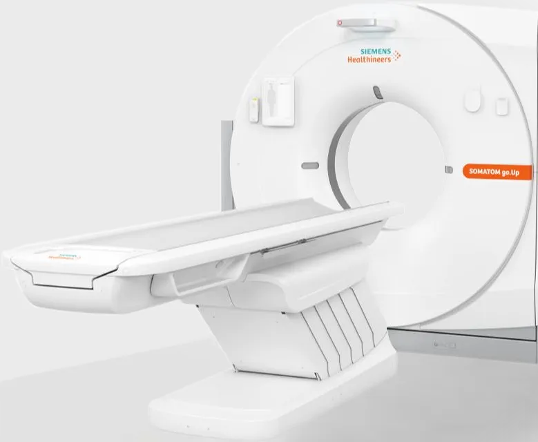
A 64-slice CT scanner is an advanced computed tomography (CT) machine that captures multiple high-resolution images of your internal organs and structures simultaneously. The term "64-slice" refers to the number of image "slices" the scanner can obtain in a single rotation, allowing for rapid image acquisition and detailed 3D reconstructions.
How does a 64-slice CT scanner differ from a traditional CT scanner?
A 64-slice CT scanner captures more images in a single rotation than traditional CT scanners, which typically have 16 or fewer slices. This increased imaging capacity allows for faster scan times, higher resolution images, and reduced radiation exposure for patients.
Is a 64-slice CT scanner safe?
Yes, a 64-slice CT scanner is safe for most patients. However, CT scans do use ionizing radiation to produce images, so it's essential to weigh the potential risks and benefits of the procedure. At Neuro Imaging Winter Park, our highly trained staff takes all necessary precautions to minimize radiation exposure and ensure patient safety.
What types of conditions can a 64-slice CT scanner diagnose?
A 64-slice CT scanner can diagnose a wide range of medical conditions, including cancer, heart disease, vascular disorders, and trauma. The high-quality images produced by the scanner provide essential diagnostic information and help guide treatment planning.


What is MR Arthrography?
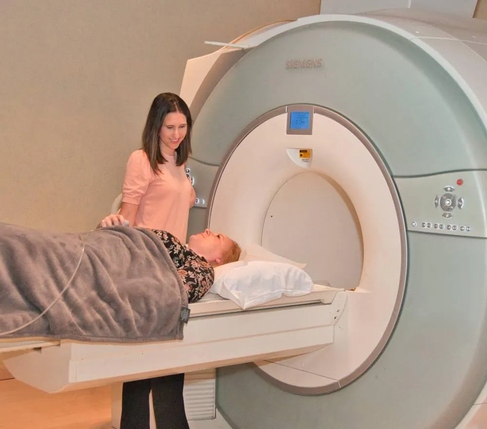
MR Arthrography is a two-step diagnostic imaging procedure that involves the injection of a contrast agent into a joint, followed by an MRI scan. The contrast agent helps to outline the joint structures, such as ligaments, tendons, and cartilage, providing clearer and more detailed images than a traditional MRI.
When is MR Arthrography used?
MR Arthrography is used when traditional MRI may not provide sufficient detail to diagnose certain joint conditions. This advanced imaging technique is particularly useful for evaluating:
Joint abnormalities, such as labral tears in the hip or shoulder
Cartilage injuries or degeneration in the knee, ankle, or wrist
Ligament and tendon injuries or inflammation
Suspected joint infections

Advantages of MR Arthrography:
Enhanced Visualization
The use of a contrast agent during MR Arthrography improves the visualization of joint structures, enabling more accurate diagnosis of joint abnormalities.
Comprehensive Evaluation
MR Arthrography allows for a detailed assessment of joint structures, including ligaments, tendons, and cartilage, helping physicians identify the cause of joint pain or dysfunction.
Minimally Invasive
Although MR Arthrography requires the injection of a contrast agent into the joint, it is a minimally invasive procedure that typically causes minimal discomfort.
Guided Treatment Planning
The detailed images obtained through MR Arthrography can help guide treatment planning and ensure appropriate management of joint conditions.
Advantages of Using 3T MRI:
High-Resolution Images:
The 64-slice CT scanner produces exceptionally detailed images, allowing for improved visualization of internal structures and enhanced diagnostic capabilities.
Faster Scan Times:
With the ability to capture more images per rotation, the 64-slice CT scanner significantly reduces scan times, resulting in increased patient comfort and convenience.
Lower Radiation Exposure:
The advanced technology of the 64-slice CT scanner enables efficient imaging with reduced radiation exposure, minimizing potential risks for patients.
Advanced 3D Reconstruction:
The 64-slice CT scanner allows for sophisticated 3D reconstruction of images, providing better visualization and planning for physicians, particularly in complex cases.
Choose Neuro Imaging Winter Park
For your MR Arthrography needs and experience the difference that advanced technology and expert care can make in your diagnostic journey. Schedule your appointment today and take the first step towards improved joint health.


Our Doctors
Healthia’s team of qualified doctors is always ready to help you.
Location & Directions
Our clinic is situated downtown and is always available for you.
Appointments
We recommend scheduling an appointment in advance.
IMAGING SERVICES

Anesthesiology & Pain Management

Bariatric &
Metabolic Institute

Cancer Center
Why Neuro Imaging Winter Park?

Qualified Doctors
We employ recognized medical specialists from all over the world.

Modern Equipment
Our team uses the newest medical equipment and supplies of top quality.

Emergency Help
Our qualified specialists are always ready to provide any emergency help.

Individual Approach
Our unique approach allows us to provide tailored medical services.
Our Doctors
The Right Diagnosis
Your care is our top priority. Our wide, short-bore 32 channel 3T MRI offers exceptional patient comfort. Our expert doctors and advanced technology provide superior quality imaging for all of our patients.
© 2024 All rights reserved.


