
Neuro Imaging Winter Park
The center with the ultimate quality MRI and CT services, where all patients are treated as VIPs.
Experience the difference with our physician-owned facility, offering cost-effective diagnostic imaging and personalized care for our patients.
Neuro Imaging Winter Park
The center with the ultimate quality MRI and CT services, where all patients are treated as VIPs.
Experience the difference with our doctor-owned facility, offering cost-effective diagnostic imaging and personalized care for our patients.

Our board-certified radiologists are ready to help

Conveniently located and accessible

Click here to request an appointment
The Neuro Imaging Difference
Welcome to Neuro Imaging Winter Park, the premier provider of MRI and CT scan services in Winter Park, FL. Our state-of-the-art,
physician-owned facility guarantees you receive top-notch care and exceptional services at affordable prices. By choosing our center, you can significantly compare to hospital imaging departments without compromising on the quality of your diagnosis.
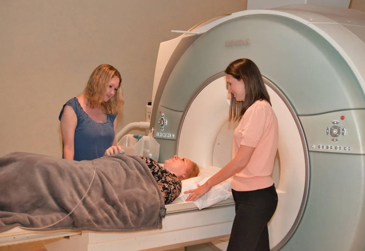
Our advanced imaging technology includes the powerful 32 Channel 3T MRI machine and the efficient 64-slice CT scanner, both designed to provide accurate and detailed images in a timely manner. Trust our team of experienced professionals to help you on your journey to better health.
Why Choose Neuro Imaging Winter Park:
Physician-owned and operated, ensuring personalized care and attention
Cutting-edge imaging technology for accurate and timely diagnosis
A compassionate and professional team dedicated to your well-being
Affordable pricing without sacrificing quality
Flexible scheduling options and convenient location

We have been at the forefront in Brain, Spine, Head & Neck Imaging in Central Florida for over 20 years.
What Our Patients Say
"Years ago I had this done at another place, and my wife and I were both dreading this one.. What a difference. The staff was great at explaining what was going to happen, and positioning me comfortably. If you need this procedure done.. GO HERE… Rave review"
James M.
"Five stars, They made my mom feel like family. Everyone in the office was extremely friendly and caring. We will not be going anywhere else for a scan. Highly Recommend! 😊"
Stacy P.
"I’ve been to this office 2 times now for MRI exams. The MRI tech is great and really knows what he’s doing. If I need imaging in the future, I’ll definitely go back to this place!"
David L.
Why Choose Neuro Imaging Winter Park:
Doctor-owned and operated, ensuring personalized care and attention
Cutting-edge imaging technology for accurate and timely diagnosis
A compassionate and professional team dedicated to your well-being
Affordable pricing without sacrificing quality
Flexible scheduling options and convenient location
We have been at the forefront in Brain, Spine, Head & Neck Imaging in Central Florida for over 20 years.
Meet Our Team
Neuro Imaging Winter Park is proud to announce Dr. Rajendra Vazirani, who is an expert in musculoskeletal, body, and oncologic imaging, is now a full partner at our practice. With Dr. Vazirani's added expertise, our patients receive top quality scans in musculoskeletal, body, and oncologic imaging comparable to what they would receive at the best academic institutions.
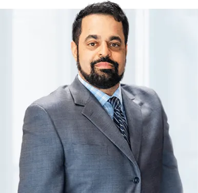
Rajendra M. Vazirani, M.D.
ONCOLOGIC RADIOLOGY • BODY AND MSK RADIOLOGY • BOARD CERTIFIED ABR

Marc D.
Shapiro, M.D.
NEURORADIOLOGY • BOARD CERTIFIED ABR • CAQ NEURORADIOLOGY
Our Doctors
Healthia’s team of qualified doctors is always ready to help you.
Location & Directions
Our clinic is situated downtown and is always available for you.
Appointments
We recommend scheduling an appointment in advance.
IMAGING SERVICES

Anesthesiology & Pain Management

Bariatric &
Metabolic Institute

Cancer Center
Why Neuro Imaging Winter Park?

Qualified Doctors
We employ recognized medical specialists from all over the world.

Modern Equipment
Our team uses the newest medical equipment and supplies of top quality.

Emergency Help
Our qualified specialists are always ready to provide any emergency help.

Individual Approach
Our unique approach allows us to provide tailored medical services.
Our Doctors


The Right Diagnosis
Your care is our top priority. Our wide, short-bore 32 channel 3T MRI offers exceptional patient comfort. Our expert doctors and advanced technology provide superior quality imaging for all of our patients.
© 2024 All rights reserved.